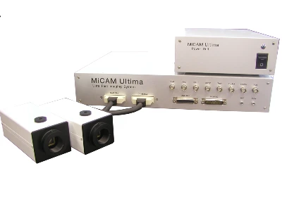
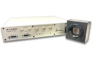
MiCAM ULTIMA, is a CMOS based ultra high speed imaging system that optimizes high speeds, high resolution, and high signal to noise ratios. It is designed to detect very small changes in fluorescent light levels of biological samples that are stained with fluorescence probes such as voltage-sensitive dyes or calcium indicators.
MiCAM ULTIMA
* In 14 years, 114 units of MiCAM ULTIMA were sold and production has already
ended.
* Next model MiCAM03-N256 is now available.
* For software download, go to the "Download" page.
* Next model MiCAM03-N256 is now available.
* For software download, go to the "Download" page.
Main Features
- Spatial Resolution
- 100 x 100 pixels
- Image Sensor Size
- 10mm x 10mm
- Camera Port
- 2
- Maximum Frame Rate
- 10,000fps (100x100 pixels)
Applications
- Membrane potential (voltage sensitive dye) imaging
- Calcium imaging for in vivo brain/cardiomyocytes
- Imaging with FRET, GCaMP, GEVI
- Intrinsic optical signal imaging based on hemoglobin and flavoprotein autofluorescence
- Ratiometric fluorescence imaging with 2 camera heads
- Panoramic imaging with 2 camera heads
- Other high speed imaging
Features
Maximum Frame Rate of 0.1msec/frame (10KHz)
 In order to record small changes in fluorescence of biological samples
stained with voltage sensitive dyes, the ideal combination of high
sensitivity and high speed is required. MiCAM ULTIMA, which overcomes
these challenges with BrainVision's cutting-edge research and
development, can detect small changes in fluorescence such as 0.1% After
acquisitions, users can easily analyze signal propagation between
neurons of samples such as hippocampus slices at speeds up to 10,000
frames per second and only 4 times averaging.
In order to record small changes in fluorescence of biological samples
stained with voltage sensitive dyes, the ideal combination of high
sensitivity and high speed is required. MiCAM ULTIMA, which overcomes
these challenges with BrainVision's cutting-edge research and
development, can detect small changes in fluorescence such as 0.1% After
acquisitions, users can easily analyze signal propagation between
neurons of samples such as hippocampus slices at speeds up to 10,000
frames per second and only 4 times averaging.
Real-Time Imaging of Action Potential Propagation with Single Scan
 Temporal resolution of 0.1msec/frame, spatial resolution of
100x100pixel, and dynamic range of 70db makes it possible for scientists
to record neuronal activity at speeds of several millisecond/frame even
without any averaging acquisition (single scan).
Temporal resolution of 0.1msec/frame, spatial resolution of
100x100pixel, and dynamic range of 70db makes it possible for scientists
to record neuronal activity at speeds of several millisecond/frame even
without any averaging acquisition (single scan).
Huge Well Depth of Single Pixel (10Me- / 1.5Me-)
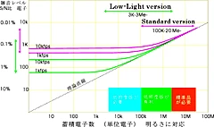 Sufficient fluorescence light intensity is needed to detect small
changes in fluorescence with a single sweep. MiCAM ULTIMA, which has a
huge well depth (10Me- / 1.5Me-), can achieve high S/N ratios without
any pixel saturation in most bright light conditions.
Sufficient fluorescence light intensity is needed to detect small
changes in fluorescence with a single sweep. MiCAM ULTIMA, which has a
huge well depth (10Me- / 1.5Me-), can achieve high S/N ratios without
any pixel saturation in most bright light conditions.
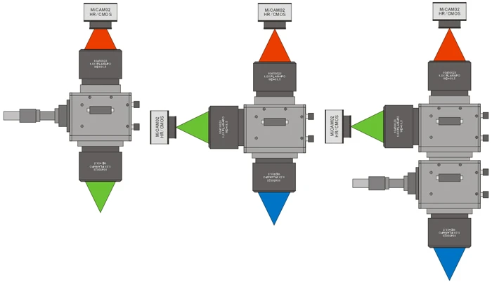
Completely Synchronized Dual Camera System
MiCAM ULTIMA processor has two camera ports. The ULTIMA can operate as a synchronized dual camera system by the simple addition of a slave camera and connection to the existing processor. This allows for dual-wavelength imaging or dual-staining with multiple probes such as voltage-sensitive dyes and calcium indicators.
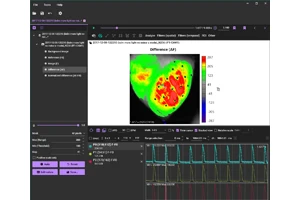
Software with Intuitive Interface
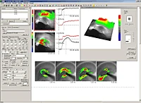 BV_Analyzer is a data analysis software and is included with all of the
MiCAM systems. This software has various functions, such as wave
display, 3D display, animation display, real-image display,
spatial/temporal filter, high-pass/low-pass filter, AVI/TIFF/BMP/CSV
file export, etc.
BV_Analyzer is a data analysis software and is included with all of the
MiCAM systems. This software has various functions, such as wave
display, 3D display, animation display, real-image display,
spatial/temporal filter, high-pass/low-pass filter, AVI/TIFF/BMP/CSV
file export, etc.
Characteristics of Wave Spectral Sensitivity
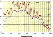 This sensor has a wide range of spectral sensitivity between near-infra
red (800-900nm) to ultraviolet (300-400nm), and has a high quantum
efficiency of 50-60%. As a result, the ULTIMA is suitable for various
imaging techniques with many different kinds of fluorescent dyes.
This sensor has a wide range of spectral sensitivity between near-infra
red (800-900nm) to ultraviolet (300-400nm), and has a high quantum
efficiency of 50-60%. As a result, the ULTIMA is suitable for various
imaging techniques with many different kinds of fluorescent dyes.
Sample data
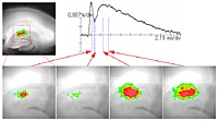
Rat Slice Imaging at 10KHz
Action potential propagation was recorded at 0.1msec/frame. 10kHz recording allows us to identify presynaptic activity and postsynaptic activity separately.
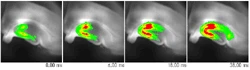
"Mouse In Vivo Imaging using Voltage-Sensitive Dye
This data was obtained from a 2 month-old mouse that was under urethane anaesthesia (1.75 g/kg). A heating pad was used to maintain the measured body temperature of the mouse at 37°C. Two electrodes were inserted under the skin of the forearms to receive the electrocardiogram signal which was used to trigger the MiCAM ULTIMA acquisition.
Specifications
| Model | MiCAM ULTIMA | |
|---|---|---|
| ULTIMA-H | ULTIMA-L | |
| Sensor | Original CMOS | |
| Actual Sensor Size | 10mm x 10mm | |
| A/D Converter | 14bit (Data resolution is 16 bit after digital process) | |
| Spatial Resolution (pixels) | 100(H) x 100(V) | |
| Temporal Resolution (/frame) | 0.1-250.0ms | |
| Maximum Frame Number | 20,480 (Option: 40,000) | |
| Well-Depth | 10,000,000e (Option: 24,000,000e) | 1,500,000e (Option: 3,000,000e) |
| Actual Quantum Efficiency | 63%@550nm | |
| 45%@700nm | ||
| Dark Noise | 920e@0.1msec | 230e@0.1msec |
| 400e@1.0msec | 80e@1.0msec | |
| Included | Processor, Cameras, Computer, Monitor, Acquisition/Analysis software | |
| Output/Input | 2 analog inputs (6 additional channels available), 8 digital outputs, 8 digital inputs, timing output, programmable pulse sequencer output, live display monitor output, synchro output | |
| Acquisition Software | Trigger signal output, Focus monitoring, Pre-trigger recording, Multi-stimulus output, Frame rate setting, Frame number setting, Real-image display, Master/Slave mode, Shutter controlling pulse, Auto-reference function, Auto background image recording and so on | |
| Analysis Software | dF/F, Averaging, Addition, Division, Spatial filter, Cubic filter, Temporal filter, Bleaching compensation, Vibration compensation, High-pass/Low-pass filter, Reference frame setting, 3D display, Wave display, ROI wave display, Movie, Multi image display, Real image display, AVI/TIFF/BMP/CSV output and so on | |
Optional parts
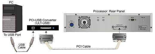
PCI-USB Interface Converter for MiCAM ULTIMA (ULT-USB)
 Previously, only the PCI card was available to connect MiCAM ULTIMA to
PCs. As newer PCs were released which used newer generations of
chipsets, incompatibility issues occurred between the PCI cards and the
newer PCs.
Previously, only the PCI card was available to connect MiCAM ULTIMA to
PCs. As newer PCs were released which used newer generations of
chipsets, incompatibility issues occurred between the PCI cards and the
newer PCs.
To avoid these compatibility issues, the PCI-USB Converter was developed. With the PCI-USB Converter, users can select any PC to use with MiCAM ULTIMA, as long as it has a USB port. The converter is compatible with Windows XP PCs, Windows 7 (32bit and 64bit) PCs, Windows 10 (64bit) PCs and Windows 11 PCs.
Next Generation of MiCAM ULTIMA
- High Speed Imaging System
- MiCAM03-N256
MiCAM03-N256 is a high-speed imaging system that captures and visualizes small changes in fluorescence intensity from biological samples stained with fluorescent probes, such as voltage sensitive dyes and calcium dyes.
- Spatial Resolution :
-
128x128 - 256x256 pixles
(32x32 pixels - 256x256 pixles with option) - Maximum Frame Rate :
- 1,000fps
(20,000 fps available with option) - Up to 2 camera heads can be used with completely synchronization.
Wide-field Imaging with High N.A
- Fluorescence Mesoscope
- THT Series
The THT is a fluorescence widefield mesoscope developed for detecting low light fluorescence that is emitted from biological sample at a low magnicifation. This mesoscope has a very simple optical path combining two large-diameter objective lenses and a custom-made large fluorescent filter