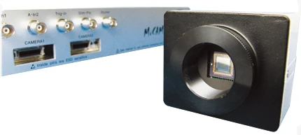
MiCAM02-CMOS is a CMOS camera that is compatible with the MiCAM02 imaging system. The originally designed CMOS sensor achieves higher frame rates and a wider dynamic range than the current CCD models. The new CMOS camera provides high resolution movies at the sub-millisecond level and creates significant advantages in high speed imaging applications.
MiCAM02-CMOS
* MiCAM02 system was discontinued. Next model
MiCAM03-N256
is now available.
* Camera head for MiCAM02 (02-HR, CMOS) is still available.
* Camera head for MiCAM02 (02-HR, CMOS) is still available.
Main Features
- Spatial Resolution
- 92 x 80 pixels / 188 x 160 pixels
- Image Sensor Size
- 5.76mm x 4.8mm
- Maximum Frame Rate
-
1,666fps (92 x 80 pixels)
833fps (188x160 pixels)
Applications
- Membrane potential (voltage sensitive dye) imaging
- Calcium imaging for in vivo brain/cardiomyocytes
- Imaging with FRET, GCaMP, GEVI
- Intrinsic optical signal imaging based on hemoglobin and flavoprotein autofluorescence
- Ratiometric fluorescence imaging with 2 camera heads
- Panoramic imaging with 2 camera heads
- Other high speed imaging
Features
Original CMOS image sensor developed by Brainvision Inc.
Higher frame rate (max: 0.6msec/frame) and lower noise
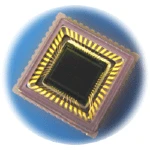
MiCAM02-CMOS sensor
The original CMOS image sensor adapted for the MiCAM02-CMOS camera is
designed by Brainvision Inc. To achieve signal readout with higher speed
and lower noise, many parts from pixel to readout circuit are tuned up
with the latest CMOS technology. This allows the CMOS camera to record
at higher frame rates of up to 0.6msec/frame at 92x80 pixels, and
1.2msec/frame at 188x160 pixels.
Wide dynamic range over 68dB
The highly customized CMOS sensor has a well depth of 450,000e- and wide dynamic range of 68dB or more. With sufficient light intensity emitted from a biological sample, it is possible to achieve higher S/N ratios than MiCAM02-HR and MiCAM02-HS.
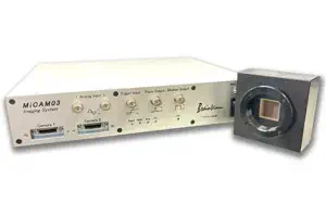
Compatible with MiCAM02
Current MiCAM02 users can easily utilize the new MiCAM02-CMOS camera by simply connecting the camera to the processor and updating the software version.
Synchronized dual-camera system for dual-wave length imaging
Two CMOS cameras can be connected to the MiCAM02 processor to do synchronized recordings. This dual-camera system can be used to simultaneously image voltage-sensitive dye and calcium ion indicators, as well as perform 3D mapping of multiple locations on a biological sample.
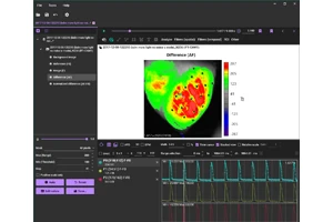
Sample Data
Imaging of Action Potential Propagation in Isolated Rat Heart
An action potential propagation in isolated rat heart. The heart was Langendorff perfused, stained with Di-4-ANEPSS, and recorded at 1,000 frames/second and 92x80 pixels using MiCAM02-CMOS camera.
Specifications
| Process Technology | 0.18CIS-P1M6 |
|---|---|
| Resolution (pixels) | 188(H) x 160(V) / 92(H) x 80(V) |
| Pixel Size | 30 μm square |
| Pixel Aperture Ratio | 63% |
| Well Depth | 450,000 e- |
| Readout Noise | < 150e- |
| Dynamic Range | > 68dB |
| Temporal Noise Factor | < -57dB |
| Temporal Noise Factor | < -57dB |
| Chip Size | 7.0mm square |
| Max. Frame Rate | 1,700 fps |
| Compatibility | MiCAM02 |
| Shutter | Original Global Shutter |
Next Generation of MiCAM02
- High Speed Imaging System
- MiCAM03-N256
MiCAM03-N256 is a high-speed imaging system that captures and visualizes small changes in fluorescence intensity from biological samples stained with fluorescent probes, such as voltage sensitive dyes and calcium dyes.
- Spatial Resolution :
-
128x128 - 256x256 pixles
(32x32 pixels - 256x256 pixles with option) - Maximum Frame Rate :
- 1,000fps
(20,000 fps available with option) - Up to 2 camera heads can be used with completely synchronization.