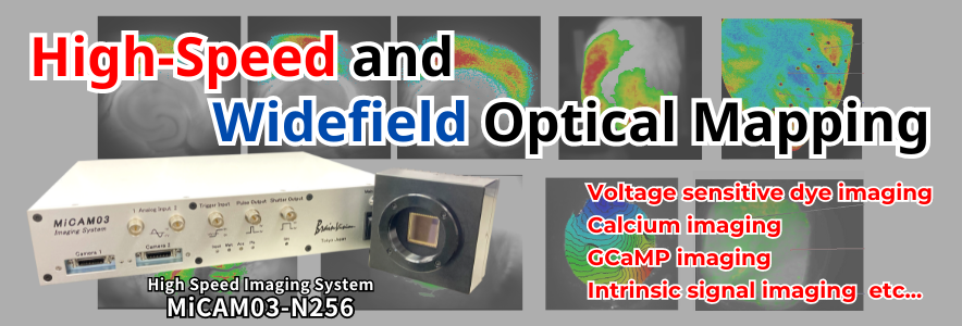
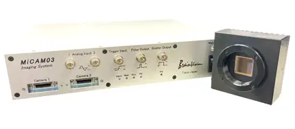
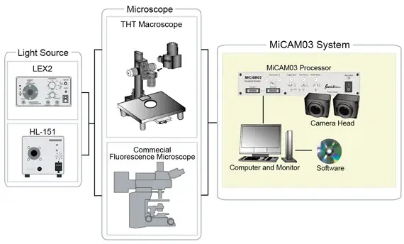
High-Speed Optical Mapping System MiCAM03-N256
MiCAM03-N256 is a high-speed optical mapping system that captures and visualizes small changes in fluorescence intensity from biological samples stained with fluorescent probes, such as voltage sensitive dyes and calcium dyes.
This product is for research use only (RUO).
Applications
- Membrane potential (voltage sensitive dye) imaging
- Calcium imaging for in vivo brain/cardiomyocytes
- Imaging with FRET, GCaMP, GEVI
- Intrinsic optical signal imaging based on hemoglobin and flavoprotein autofluorescence
- Ratiometric fluorescence imaging with 2 camera heads
- Panoramic imaging with 2 camera heads
- Other high speed imaging
Main Features
- Spatial Resolution
-
256 x 256 pixles, 128 x 128 pixels
(Optional license allows to select more pixel settings) - Image Sensor Size
- 17.6mm x 17.6mm
- S/N Ratio
- > 71dB
- Camera Port
- 2
- Memory
- 1GB
- Maximum Frame Rate
-
1,000fps (256 x 256px), 1,000fps (128 x 128px)
(Optional license allows to select faster frame rates)
Sample Data
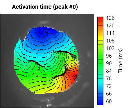
Isolated Pig Heart 1,818fps at 256x256 pixels
- Sample
- Isolated pig heart
- Fluorescence Dye
- Voltage sensitive dye
- Mapping System
- MiCAM03-N256
- Pixels
- 256x256
- Frame Rate
- 1,818fps (0.55msec/frame)
- Provided by
- Dr. Jack M. Rogers, The University of Alabama at Birmingham
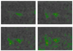
Monolayer of neonatal mouse ventricular myocyte stained with voltage sensitive dye
- Sample
- Monolayer of neonatal mouse ventricular myocyte. Sample was scratched with a needle tip at three locations (visible on the fluorescence pictures) to induce conduction blocks and meandering of the wavefront.
- Method
- Sample is paced locally at different frequencies using a glass microelectrode positioned at the edge of the sample
- Fluorescence Dye
- Voltage sensitive dye (Di-8-Anepps)
- Mapping System
- MiCAM03-N256
- Pixels
- 256x256
- Frame Rate
- 1,000fps (1.0msec/frame)
- Provided by
- Dr. Jan Kucera and Dr. Ange Maguy, Department of Physiology, University of Bern
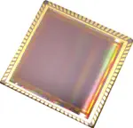
Features : Camera Head
N256 camera head is equipped with a new CMOS image sensor designed and developed by Brainvision Inc.
Large Sensor and Large Pixel
- Active Area size
- 17.6mm x 17.6mm
- Pixel size
- 69μm x 69μm
 Applications such as membrane potential imaging require both very high
speed, and the ability to detect small changes in fluorescence, such as
0.5-1%. In order to optimize S/N ratios, it is important to increase the
amount of light captured by one pixel. It is easy to collect light and
capture small changes using a sensor having large pixels.
Applications such as membrane potential imaging require both very high
speed, and the ability to detect small changes in fluorescence, such as
0.5-1%. In order to optimize S/N ratios, it is important to increase the
amount of light captured by one pixel. It is easy to collect light and
capture small changes using a sensor having large pixels.
High Spatial Resolution, High Frame Rates, and High S/N Ratios
- Number of Pixels
- 256 x 256 pixels
- Maximum Frame Rate
- 1,000 fps (at 256x256 pixels)
- Dynamic Range
- 71dB
The overall performance has been improved in a balanced manner. This includes maximum frame rate, spatial resolution, and S/N ratios, all of which are important factors for high-speed membrane potential imaging.
Wide Dynamic Range Mode or High Sensitivity Mode by Adjustable Well Depth
- Wide Dynamic Range Mode
- 3,000,000 electrons
- High Sensitivity Mode
- 600,000 electrons
It is now possible to easily switch the full well capacity of the camera
in the acquisition software. Two modes are available: wide dynamic range
mode (D-mode), and high sensitivity mode (S-mode).
D-mode is
used for tissue samples with high fluorescence light intensity, and
S-mode is used for cultured cell monolayers, or preps with low
fluorescence light intensity. This adjustable well depth makes it
possible to image a variety of biological samples using a single
camera.
The well-depth is fixed regardless of settings such as sampling rate.
ROI Readout for Higher Frame Rates (Option)
Preset partial readout setting and multiple ROI setting can be selected, and as a result it is possible to achieve higher frame rates. For example, ultra high frame rates such 20,000 fps@32x32 pixels is possible.
-
 All pixels readout (256x256 pixels)
-
 Multiple ROI readout (32x32x9 pixels)
Features : Processor
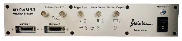
Direct Data Saving and Long-Term Data Acquisition with USB3.0 High-Speed Data Transfer
New USB3.0 interface allows for significantly faster data transfer from processor to PC. It is now possible to save data directly to HDD or SSD, and long-term recording of several minutes up to a few hours can be achieved, regardless of the RAM capacity (Note that sampling rate, number of pixels, number of cameras used, and PC specifications, will affect the total recording time).
| Disk Capacity | Pixels | Frame Rate (fps) | Number of recordable frames | Recordable time (min) |
|---|---|---|---|---|
| SSD 1TB | 256x256 | 500 | 6,840,000 | 228.0 |
| 1,000 | 6,840,000 | 114.0 | ||
| 1,818 | 6,840,000 | 62.7 | ||
| 128x128 | 500 | 27,360,000 | 912.0 | |
| 1,000 | 27,360,000 | 456.0 | ||
| 5,263 | 27,360,000 | 86.6 |
(*Approximate number of recordable frames and recordable time shown above are averaged values that were measured using the PC configuration which is included with MiCAM03. There is no guarantee that the results will be the same in all circumstances.)
Up to two cameras can easily be connected and used in a completely synchronized dual-camera system
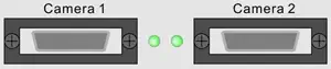
MiCAM03 processor has two camera ports, and can be adapted as a dual-camera system using an optional second camera, which enables synchronized recordings. This allows for simultaneous imaging of multiple fluorescent wavelengths, as well as three-dimensional imaging from different angles.
Synchronous Lighting Control of LED Light Source for Two-Wavelength Excitation Imaging such as GCaMP Imaging
In the case of imaging using two fluorescent probes with two different
excitation wavelengths or imaging using a two-wavelength
excitation/single-wavelength fluorescent probe, it is necessary to
synchronize the frame timing of the camera and the lighting timing of
the light source.
This function is to output the lighting signal synchronized with each
frame timing from MiCAM03 to multiple LED light sources. This makes it
possible to easily synchronize the frame acquisition with MiCAM03 and
LED lighting.
There are up to 4 channels of output. Preset patterns, in which signals
are output alternately from each channel, are available. There is no
need to set the lighting pattern using a commercially available pulse
generator.
Moreover, since the pulse delay time and pulse width can be specified by
software, it is possible to make detailed settings in accordance with
the rise and fall times of the LED used.
-
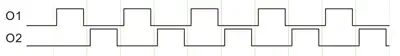 Two-wavelength excitation mode
Two-wavelength excitation for GCaMP imaging using LEX9 and MiCAM
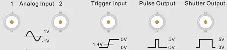
Various I/O Connectors, and Improved Compatibility with External Devices
MiCAM03 has multiple input and output connections available. These universally standard I/O connections can be synchronized with image acquisition and external devices.
Other Features
Turnkey System Ready for Immediate Use
The system consists of hardware/software specifically designed for high-speed fluorescence imaging, and the PC and monitor are included as standard equipment. Before use, calibration/setting/tuning is not necessary. The hardware connection is straightforward between components using cables, making it possible to acquire data immediately after setup.
Pre-Post Trigger Mode
MiCAM03 can record activity which occurs before and after a trigger signal is generated. When an external trigger occurs, such as a spontaneous biological signal, or a trigger pulse from an external stimulator, a specified amount of "pre-trigger" data can be saved. The "post-trigger" data is also collected in the same recording. As a result, both pre-trigger data and post-trigger data can be saved within one acquisition period.
-
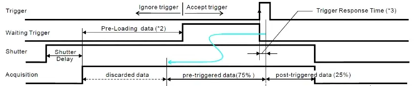 Pre-Post Trigger Mode (75% Pre-Trigger)
Time Lapse Imaging
MiCAM03 can capture phenomenon that changes in a few minutes up to several hours, intermittently. Because illumination light is turned on only at the time of image acquisition, phototoxic effects to sample is minimized, and a stable baseline can be acquired.
-
 Example of Time-lapse imaging
High Resolution Camera Control Function (Optional)
It is possible to control a high resolution camera having 1920x1200 pixels (BV-XB1 camera). High resolution imaging can be applied to various applications, including detection of intracellular Ca 2+ dynamics, flavoprotein fluorescence, FRET imaging, and also imaging of animal behaviors. High resolution imaging with BV-XB1 can also be synchronized with MiCAM03 high-speed imaging.
-
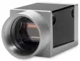 BV-XB1
-
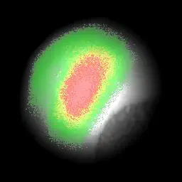
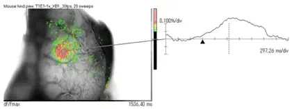 Example of flavoprotein imaging
Software
Acquisition/Analysis Software (BV Workbench)
BV Workbench is the image acquisition software that controls MiCAM03-N256 optical mapping system. In addition to image acquisition and control of inputs and outputs of the optical mapping system, analysis functions including movie playback, wave display, filter functions, etc…are also included. BV Workbench has an intuitive user interface that is easily operated with the use of a mouse.
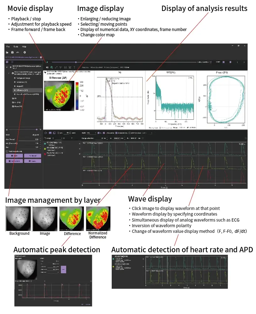
Cardiac Optical Mapping Data Anaysis using BV Workbench Version 4.5.1
Specifications
| System | ||
|---|---|---|
| Name |
High-Speed Optical Mapping System MiCAM03-N256 |
High Speed Optical Mapping System MiCAM03-HR |
| Model | MC03-N256 | MC03-HR |
| Standard System Configuration |
Camera Head (1-2), Processor, PC, Monitor, Acquisition software, Analysis Software |
|
| Supported OS | Windows 7/10 64bit | |
| Camera Head | ||
| Name | N256 camera | HR camera |
| Model | MC-N256CAM | 02HRCM |
| Image Sensor | Original CMOS | Sony CCD |
| Active Pixels (H x V) | 256 x 256 | 380 x 244 |
| Active Image Area (H x V) | 17.6mm x 17.6mm | 6.4mm x 4.8mm |
| Pixels Size (H x V) | 69μm x 69μm | 16.8μm x 19.6μm |
| Shutter Mode | Global | Global |
|
Maximum Frame Rate and Pixels (for 1 camera) |
1,923fps (256x256px) 3,125fps (192x192px) 5,556fps (128x128px) 20,000fps (32x32px) |
143fps (380x244px) 270fps (188x120px) 455fps (92x60px) |
| Quantum Efficiency | 55%@550nm / 50%@600nm | 75%@550nm / 50%@700nm |
| Well-Depth | 3,000,000e- / 600,000e- (switching) | 100,000e- (180x120px) / 12,000e- (180x120px, H-bin) |
| Dark Noise | 500e- / 130e- |
60e- (3.7ms,180x120px) / 20e- (3.7ms,180x120px,H-bin) |
| Lens mount |
Original M42 mount (flange back:14.5mm) (Option: C or F mount) |
C mount |
| Interface | Special Cable | Special Cable |
| Dimension (WxHxD) | 80mm x 80mm x 50mm | 62mm x 52mm x 46mm |
| Weight | 474g | 100g |
| Processor | ||
| Name | MiCAM03 processor | MiCAM03 processor (analog I/F) |
| Model | MC03-PRC | MC03-PRC-A |
| Camera Interface | 1 | |
| Camera Port | 2 | |
| Slot for Optional Unit | - | |
| Buffer Memory Inside Processor | 1GB | |
| Analog Inputs for Synchronous Recording |
2ch | |
| Output for Stimulation Pulse | 1ch | |
| Input for External Trigger Signal | 1ch | |
| Output for Lighting Control | 1ch | |
| Input/Output for Frame Timing | 1ch | |
|
Output for Real Time Light Intensity Monitor |
- | |
| Output for Light Intensity Control | - | |
| Current Output | - | |
| Output for Acquisition Status | - | |
| Output for Trigger Wait Status | - | |
| Digital I/O port | 1 (6 pins) | |
| Trigger Mode | Software / TTL External Input / Analog Input / Usage of Optional Unit | |
| Data Transfer Mode | USB3.0 | |
| Recording Mode | Temporary storage to PC RAM / Direct data saving to PC drive (SSD/HDD) | |
| Maximum Recordable Frame Number | Dependent on camera used and image acquisition settings | |
| Power Supply | AC100V-230V、50Hz/60Hz 150W | |
| Fuse | T2A L 250V | |
| Dimension | 300mm (W) x 260mm (D) x 60mm (H) | |
| Weight | 2.6kg | Default/Optional Function |
| Single Trial Acquisition | Default | |
|
Long Time Acquisition (direct saving to disk) |
Option (sold separately) | |
| Averaging Acquisition | Default | |
|
Multiple ROI Readout for Higher Frame Rate |
Option (sold separately) | |
| Time-Lapse Acquisition | Option (sold separately) | |
| High Resolution Camera Control | Option (sold separately) | |
| Acquisition software | Default | |
| Analysis software | Option (sold separately) | |
| SDK (for Visual C# .NET and MatLab) | Option (sold separately) | |
* This product is for research use only. * This product is made in Japan.
| Selectable Number of Pixels (HxV) | Maximum Frame Rate (fps) | |||
|---|---|---|---|---|
| NR Mode OFF |
NR Mode ON 2 Times Averaging |
NR Mode ON 4 Times Averaging |
||
| 256x256 | Full Resolution | 1,818 | 1,923 | 1,031 |
| 192x192 | Square ROI | 2,941 | 3,125 | 1,695 |
| 128x128 | 5,263 | 5,556 | 3,333 | |
| 64x64 | 12,500 | 12,500 | 8,333 | |
| 256x128 |
Horizontally Long Rectangle ROI |
3,333 | 3,448 | 1,887 |
| 256x64 | 5,882 | 5,555 | 3,226 | |
| 256x32 | 9,091 | 8,333 | 5,000 | |
| 128x256 |
Vertically Long Rectangle ROI |
3,030 | 3,333 | 1,852 |
| 64x256 | 4,348 | 5,000 | 3,125 | |
| 32x256 | 5,556 | 7,143 | 4,762 | |
| 32x32 | 16 blocks | 2,381 | 2,632 | 1,724 |
| 15 blocks | 2,500 | 2,778 | 1,818 | |
| 14 blocks | 2,703 | 2,941 | 1,961 | |
| 13 blocks | 2,857 | 3,125 | 2,083 | |
| 12 blocks | 3,125 | 3,448 | 2,273 | |
| 11 blocks | 3,333 | 3,704 | 2,439 | |
| 10 blocks | 3,704 | 4,000 | 2,703 | |
| 9 blocks | 4,000 | 4,348 | 2,941 | |
| 8 blocks | 4,545 | 5,000 | 3,226 | |
| 7 blocks | 5,000 | 5,556 | 3,704 | |
| 6 blocks | 5,556 | 6,250 | 4,167 | |
| 5 blocks | 6,667 | 7,143 | 5,000 | |
| 4 blocks | 7,692 | 8,333 | 5,882 | |
| 3 blocks | 10,000 | 11,111 | 7,692 | |
| 2 blocks | 12,500 | 14,286 | 10,000 | |
| 1 blocks | 20,000 | 20,000 | 16,667 | |
| Selectable Number of Pixels (HxV) | Maximum Frame Rate (fps) | |||
|---|---|---|---|---|
| NR Mode OFF |
NR Mode ON 2 Times Averaging |
NR Mode ON 4 Times Averaging |
||
| 256x256 | Full Resolution | 1,087 | 1,064 | 1,031 |
| 192x192 | Square ROI | 1,818 | 1,754 | 1,695 |
| 128x128 | 3,704 | 3,571 | 3,333 | |
| 64x64 | 9,091 | 8,333 | 8,333 | |
| 256x128 |
Horizontally Long Rectangle ROI |
2,041 | 1,961 | 1,887 |
| 256x64 | 3,571 | 3,226 | 3,125 | |
| 256x32 | 5,882 | 5,000 | 5,000 | |
| 128x256 |
Vertically Long Rectangle ROI |
2,000 | 1,961 | 1,852 |
| 64x256 | 3,571 | 3,448 | 3,125 | |
| 32x256 | 5,556 | 5,556 | 4,762 | |
| 32x32 | 16 blocks | 1,887 | 1,613 | 1,613 |
| 15 blocks | 2,041 | 1,724 | 1,724 | |
| 14 blocks | 2,174 | 1,852 | 1,852 | |
| 13 blocks | 2,326 | 1,961 | 1,961 | |
| 12 blocks | 2,500 | 2,128 | 2,128 | |
| 11 blocks | 2,703 | 2,326 | 2,326 | |
| 10 blocks | 2,941 | 2,500 | 2,500 | |
| 9 blocks | 3,226 | 2,778 | 2,778 | |
| 8 blocks | 3,571 | 3,125 | 3,125 | |
| 7 blocks | 4,000 | 3,448 | 3,448 | |
| 6 blocks | 4,762 | 4,000 | 4,000 | |
| 5 blocks | 5,556 | 4,762 | 4,762 | |
| 4 blocks | 6,667 | 5,556 | 5,556 | |
| 3 blocks | 8,333 | 7,143 | 7,143 | |
| 2 blocks | 11,111 | 10,000 | 10,000 | |
| 1 blocks | 16,667 | 16,667 | 16,667 | |
Brochure Download
-
High-Speed Optical Mapping System MiCAM Series (2019)
-
MiCAM05-N256 and MiCAM03-N256
- Products