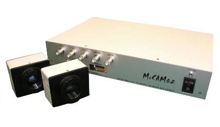
MiCAM02 has been developed as the next generation of MiCAM01 by Brainvision Inc. MiCAM01 has already achieved a high level of success among high speed imaging systems. MiCAM02 maintains the basic concepts of MiCAM01 such as high speed and high S/N ratios, but it has been significantly improved with regards to spatial resolution, sensitivity, and dark noise/read-out noise levels. Additional software functions including the ability to do time-lapse recordings, have also been implemented.
MiCAM02
* Camera head for MiCAM02 (02-HR, CMOS) is still available.
Main Features
- Spatial Resolution
- 40 x 28 pixels - 376 x 252 pixels
- Image Sensor Size
- 6.4mm x 4.8mm
- Camera Port
- 2
- Maximum Frame Rate
- 769fps (40x28 pixels)
Applications
- Membrane potential (voltage sensitive dye) imaging
- Calcium imaging for in vivo brain/cardiomyocytes
- Imaging with FRET, GCaMP, GEVI
- Intrinsic optical signal imaging based on hemoglobin and flavoprotein autofluorescence
- Ratiometric fluorescence imaging with 2 camera heads
- Panoramic imaging with 2 camera heads
- Other high speed imaging
Features
CMOS camera and CCD camera are available
Maximum frame rate is 1.7
kHz
The imaging of neuronal activity, which requires the ability to detect very weak signals, adopts averaging (or accumulation) methods in most cases. MiCAM02 can be used for most experiments by utilizing the averaging method or by recording single sweeps of activity. Both CMOS and CCD cameras are available for making imaging area sizes or frame rates suitable for a wide range of experiments.
Spatial resolution is adjustable according to frame rate
Spatial resolution of MiCAM02 is varied at 40x28 - 376x252 pixels according to the frame rate selected. This flexibility with regards to binning provides higher spatial and temporal resolution.
Powerful in dark light levels
The new "H-bin mode" function reduces dark noise, and it is comparable to that of a cooled-CCD camera. This function is very effective for situations of dark light or fluorescence.
Synchronized Dual-camera system for dual-wave length imaging
MiCAM02 processor has two camera ports, and can be adapted as a dual-camera system using an optional second camera, which enables synchronized recordings. The dual-camera system can be used for the ratio-imaging with voltage-sensitive dyes or calcium ion indicators, as well as imaging with multiple probes.
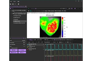
User-friendly software
Data analysis software "BV_Ana," which has many useful functions, also includes acquisition capabilities resulting in simpler, smoother, and faster experiments. The quick analysis ability of recorded data makes it possible for users to do many experiments on a single biological sample under various conditions.
Specifications
| Model | MiCAM02-CMOS | MiCAM02-HR | MiCAM02-HS |
|---|---|---|---|
| Sensor | Original CMOS | 1/2 inch CCD | 1/5 inch CCD |
| Actual Sensor Size | 5.76 x 4.8 mm2 | 6.4 x 4.8 mm2 | 2.9 x 2.1mm2 |
| Spatial Resolution (pixels) | 92(H) x 24(V) | 40(H) x 28(V) | |
| 92(H) x 40(V) | 88(H) x 60(V) | ||
| 92(H) x 80(V) | 184(H) x 124(V) | ||
| 188(H) x 160(V) | 376(H) x 252(V) | ||
| Temporal Resolution (/frame) and Spatial Resolution | 0.2ms@92x24 | 1.3ms@40x28 | 0.7ms@40x28 |
| 0.3ms@92x40 | 2.2ms@88x60 | 1.0ms@88x60 | |
| 0.6ms@92x80 | 3.7ms@184x124 | 1.8ms@184x124 | |
| 1.2ms@188x160 | 7.0ms@376x252 | 3.3ms@376x252 | |
| < 50ms | < 153ms | ||
| Maximum Recordable Frame Number | 8,720@92x24 | 21,840@40x28 | |
| 8,720@92x40 | 21,840@88x60 | ||
| 8,720@92x80 | 5,460@184x124 | ||
| 4,260@188x160 | 1,364@376x252 | ||
| Well-Depth (in H-bin mode) |
450,000e- |
100,000e-@88x60 (6,000e-@88x60) |
60,000e-@88x60 (7,500e-@88x60) |
| 100,000e-@184x124 (12,000e-@184x124) | |||
| Actual Quantum Efficiency | 45%@550nm | 75%@550nm | 50%@550nm |
| 38%@700nm | 50%@700nm | 25%@700nm | |
| Dark Noise | 150e@1.2ms, 188x160 | 40e@2.2ms, 88x60 (15e@2.2ms, 88x60) |
60e@1ms, 88x60 (25e@1ms, 88x60) |
| 60e@3.7ms, 184x124 (20e@3.7ms, 184x124) |
|||
| A/D Converter | 12bit (Data resolution is more than 14bit after digital process) | ||
| Composition | Processor, Cameras, Computer, Monitor, Acquisition/Analysis software | ||
| Trigger Output | Programmable (pulse delay, duration, pulse number, interval and so on) | ||
| Analog Inputs | 2ch (maximum 4ch with optional unit) | ||
| Other Output | 1ch | ||
| Interface | PCI (Low Profile) / PCI-Express x1 / USB | ||
| Acquisition Software | Trigger signal output, Focus monitoring, Pre-trigger recording, Multi-stimulus output, Frame rate setting, Frame number setting, Real-image display, Master/Slave mode, Shutter controlling pulse, Auto-reference function, Auto background image recording and so on | ||
| Analysis Software | dF/F, Averaging, Addition, Division, Spatial filter, Cubic filter, Temporal filter, Bleaching compensation, Vibration compensation, High-pass/Low-pass filter, Reference frame setting, 3D display, Wave display, ROI wave display, Movie, Multi image display, Real image display, AVI/TIFF/BMP/CSV output and so on | ||
Next Generation of MiCAM02
- High Speed Imaging System
- MiCAM03-N256
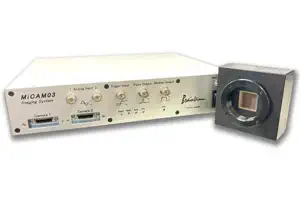
MiCAM03-N256 is a high-speed imaging system that captures and visualizes small changes in fluorescence intensity from biological samples stained with fluorescent probes, such as voltage sensitive dyes and calcium dyes.
- Spatial Resolution :
-
128x128 - 256x256 pixles
(32x32 pixels - 256x256 pixles with option) - Maximum Frame Rate :
- 1,000fps
(20,000 fps available with option) - Up to 2 camera heads can be used with completely synchronization.