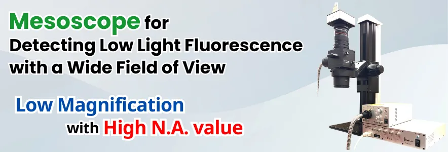
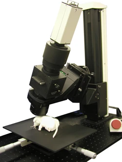
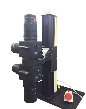
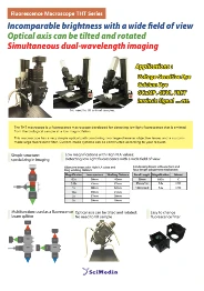
THT Mesoscope for Fluorescence Imaging
The THT is a fluorescence widefield mesoscope developed for detecting low light fluorescence that is emitted from biological sample at a low magnicifation.
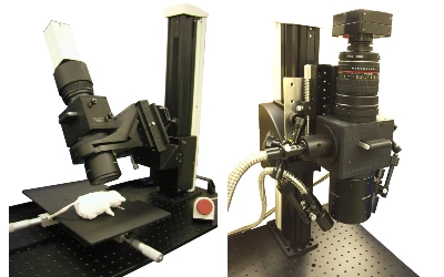
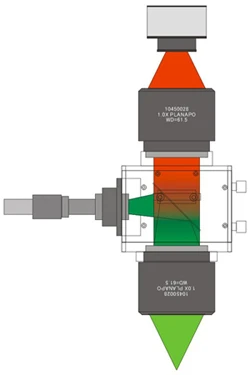
This mesoscope has a very simple optical path combining two large-diameter objective lenses and a custom-made large fluorescent filter.
Custom-made systems can be constructed according to your request.
Applications
- Voltage sensitive dye imaging
- Widefield calcium imaging for in vivo brain
- Imaging with FRET, GCaMP, GEVI
- Intrinsic optical signal imaging based on hemoglobin and flavoprotein autofluorescence
- Optogentics and imaging
- Ratiometric fluorescence imaging with multiple camera heads
Main Features
- Low magnifications with high N.A values - suitable for mesoscopic imaging
- Simple structure: specializing in imaging
- The optical axis can be tilted and rotated: no need to tilt the animal sample
- Easy removal of fluorescent filter
- Multifunction: used as a fluorescence beam splitter
- Inverted type is also possible
Features
Low Magnifications with High N.A Values:
Detecting Low Light Fluorescence with a Wide Field of View
Depending on the combination of lenses, the total magnification is about
0.19x to 6.3x (Actual field of view varies depending on the sensor size
of the camera used).
Because a large diameter and high numerical aperture lens is used,
extremely bright imaging is possible. Signals that were previously
difficult to detect may be easier to detect, and even samples that could
be captured so far may have higher S/N ratios.
Objective lens with large diameter and long working distance
|
Objective Magnifications |
Lens Diameter |
Working Distance |
|---|---|---|
| 0.3x | 50mm | 140mm |
| 0.63x | 43mm | 67mm |
| 1x | 60mm | 62mm |
| 1.6x | 59mm | 31mm |
| 2x | 57mm | 20mm |
| 5x | 39mm | 19mm |
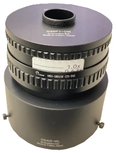
Projection lens with aperture/focal length adjustment function
|
Projection Lens |
Magnification | Mount |
|---|---|---|
| 50mm | 0.63x | C |
| 85mm/1.4 | 1.0x | EF / T / C |
| 135mm/2.0 | 1.6x | EF / T / C |
Simple Structure: Specializing in Imaging
There is no visual observation function. It is designed to focus on camera shooting with a higher S/N ratio.
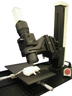
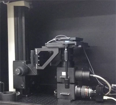
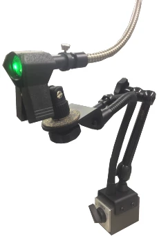
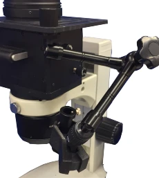
The optical axis can be tilted and rotated: no need to tilt the animal sample
A stand that can rotate the optical axis 360 degrees left and right is also selectable. By tilting the optical axis, it is possible to image the entire temporal lobe of the brain without tilting the animal sample.
Rotation
Pan
Tilt
Easy Removal of Fluorescent Filter
The fluorescent filter is held in a removable filter cube. the fluorescent filter can be changed by replacing the filter cube.
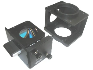
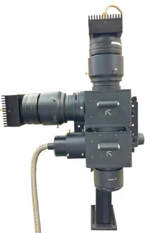
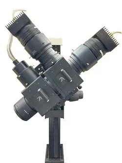
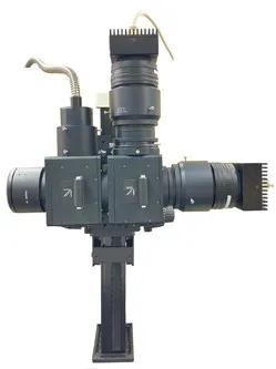
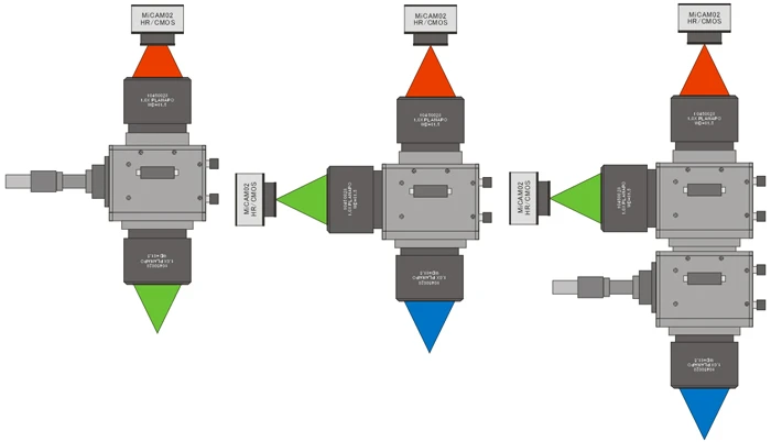
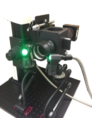
Multifunction: Used As a Fluorescence Beam Splitter
Normally, an excitation light source is connected to the left port of
the THT main unit and used as a coaxial epi-illuminator.
In addition, it can be used as a two-wavelength fluorescence splitting
device. In this case, the same lens and camera as the upper port are
connected to the left port, and two-wavelength fluorescence simultaneous
measurement using two cameras is performed.
How to Overlay Dual Camera Images
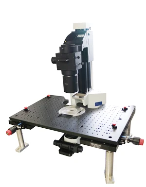
Inverted type is also possible
An inverted fluorescence mesoscope has been manufactured. The space on the sample stage becomes freer, so we expect that the degree of freedom in manipulator installation and electrode operation will increase. It can also be used with an upright fluorescent microscope. Please feel free to ask us for microscope bases and sample stages to suit the purpose of the experiment.
- THT Mesoscope
- How to Get the Best GCaMP / GEVI Signals
For bright, widefield fluorescence imaging, a mesoscope that has a
very simple design and large light path is recommended.
The
page shows 5 reasons why bright, widefield fluorescence imaging is
possible and optimized using our "THT Mesoscope".
Examples
-
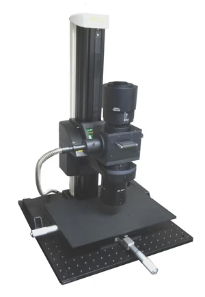
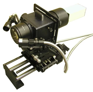
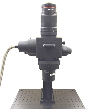 Epi illumination/single wavelength imaging
-
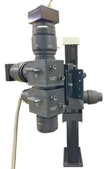
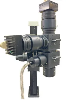
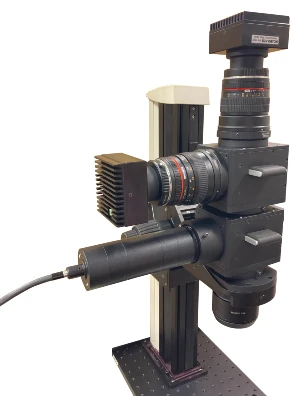
 Epi-illumination/dual wavelength imaging (upright)
-
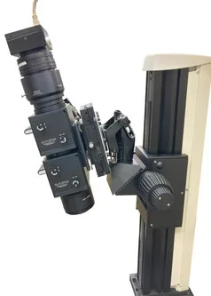
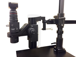
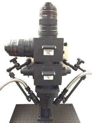
 Epi-illumination/dual wavelength imaging (45 degrees)
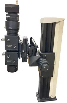
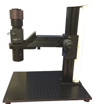
-
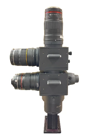
 Epi-illumination/dual wavelength imaging (90 degrees)
-
 Epi-illumination
Single wavelength imaging
Brainvision XY stage included
-
 Boom-arm stand
-
 Extension arm
-
 Olympus BX51WI base
includes manual XY table from Luigs&Neumann, Germany
-
 Horizontal imaging for Langendorff perfused heart
-
 Epi illumination
dual-wavelength imaging
-
 Triple-wavelength imaging
-
 Side illumination
dual-wavelength imaging
-
 Dual-wavelength imaging, lower position
-
 Horizontal imaging for Langendorff perfused heart
Specifications
| Name | Fluorescence Mesoscope THT Mesoscope |
|---|---|
| Optical System | Tandem lens optical system used for camera only |
| Focusing device |
Manual coarse/fine focus drive or Motorized coarse/fine focus drive (incl. dial type controller) |
| Microscope carrier | Objective nosepiece (two M65 ports), or tilting mount |
| Filter Cube |
Removable One 50mm diameter filter can be installed on the top and side Dichroic mirror mount with 2-axis tilt adjustment mechanism |
| Illumination Light for Excitation | Coaxial epi-illumination or side illumination |
| Lens port | Top: 1, Side: 1, Bottom: 1 |
| Objective lens |
PLAN 0.3x PLAN APO 0.63x PLAN APO 1x PLAN APO 1.6x PLAN APO 2x PLAN APO 5x |
| Projection lens |
PLAN APO 0.63x PLAN APO 1x PLAN APO 1.6x PLAN APO 2x 135mm/2.0 lens (w/ aperture and focus adjustment function) 85mm/1.4 lens (w/ aperture and focus adjustment function) 50mm/0.95 lens(w/ aperture and focus adjustment function) |
Approximate magnification and field size for N256 camera and THT Mesoscope
| Objective Lens | Condensing Lens (Camera Side) | |||||
|---|---|---|---|---|---|---|
| 0.63x | 1x | 1.6x | 50mm (∞ setting) |
85mm (∞ setting) |
135mm (∞ setting) |
|
| 0.3x (WD:141mm) |
0.48x (36.6mmx36.6mm) |
0.3x (58.6mmx58.6mm) |
0.19x (92.6mmx92.6mm) |
- | - | - |
| 0.63x (WD:67mm) |
1x (17.6mmx17.6mm) |
0.63x (27.9mmx27.9mm) |
0.4x (44mmx44mm) |
0.4x (44mmx44mm) |
0.69x (25.5mmx25.5mm) |
1.27x (13.9mmx13.9mm) |
| 1x (WD:61.5mm) |
1.6x (11mmx11mm) |
1x (17.6mmx17.6mm) |
0.63x (27.9mmx27.9mm) |
0.64x (27.5mmx27.5mm) |
- | 2x (8.8mmx8.8mm) |
| 1.6x (WD:30.5mm) |
2.6x (6.7mmx6.7mm) |
1.6x (11mmx11mm) |
1x (17.6mmx17.6mm) |
1.02x (17.6mmx17.6mm) |
- | - |
| 2x (WD:20.1mm) |
3.2x (5.5mmx5.5mm) |
2x (8.8mmx8.8mm) |
1.26x (13.9mmx13.9mm) |
1.28x (13.7mmx13.7mm) |
2.23x (7.9mmx7.9mm) |
4x (4.4mmx4.4mm) |
| 5x (WD:19mm) |
6.3x (2.8mmx2.8mm) |
4x (4.4mmx4.4mm) |
2.5x (7mmx7mm) |
- | 4.48x (3.9mmx3.9mm) |
- |
Reference papers for neuronal imaging
Neuronal Imaging
- Cortical State Fluctuations during Sensory Decision Making.
- Functional Differentiation of Mouse Visual Cortical Areas Depends upon Early Binocular Experience.
- Cellular and Widefield Imaging of Sound Frequency Organization in Primary and Higher Order Fields of the Mouse Auditory Cortex.
Cardiac Imaging
- Whole-heart multiparametric optical imaging reveals sex-dependent heterogeneity in cAMP signaling and repolarization kinetics.
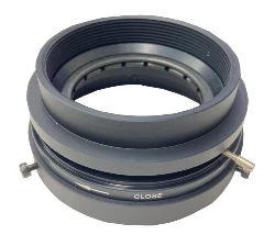
Optional parts
Lens adaptor
-
 CDAD-HL Lens Adaptor (Code No.: CDAD-HL)
Iris
-
 Imaging lens base with iris (Code No.: SM3D50-PRJM65)
Light guide arm
-
 Light Guide Holder
-
 Light Guide Arm
Flyer Download
-
Fluorescence Macroscope
THT Mesoscope