Imaging Data | Isolated heart / Cultured Cardiomyocytes
Cardiac Spheroids : Simultaneous Optical Mapping of Membrane Potential and Calcium Transients
- Sample
- Human iPS cell-derived cardiac spheroids
- Method
- Electrical Stimulation
- Fluorescence Probes
-
Voltage sensitive dye (RH237, left image)
Genetically Encoded Calcium Indicator (jRCaMP1b, right image) - Imaging System
- MiCAM03-N256 Dual Camera System
- Pixels
- 256x256
- Frame Rate
- 500fps (2.0msec/frame)
- Data provided by
-
Dr. Hanyu Zhang, Dr. Bijay Guragain, Dr. Jianyi Zhang,
and Dr. Jack M. Rogers
Department of Biomedical Engineering
The University of Alabama at Birmingham - Reference paper
-
Optogenetic stimulation and simultaneous optical mapping of
membrane potential and calcium transients in human
engineered cardiac spheroids
Journal of Molecular and Cellular Cardiology 199 (2025) 51-59
Cardiac Optical Mapping Data Analysis for hiPSC-CMs Stained with Voltage-Sensitive Dye
- Sample
- Human induced pluripotent stem cell-derived cardiomyocytes (hiPSC-CMs)
- Fluorescence Dye
- Voltage sensitive dye (FluoVolt)
- Imaging System
- MiCAM05-Ultima
- Pixels
- 100x100
- Frame Rate
- 200fps (5.0msec/frame)
- Data provided by
-
Dr. Hee Jae Jang, Dr. Vladislav Leonov and Dr. Alexey Glukhov
University of Wisconsin-Madison
Voltage Sensitive Dye Imaging of Isolated Langendorff Pig Heart at 1,818fps
- Sample
- Isolated Langendorff Pig Heart
- Fluorescence Dye
- Voltage Sensitive Dye
- Imaging System
- MiCAM03-N256
- Pixels
- 256x256
- Frame Rate
- 1,818fps (0.55msec/frame)
- Data provided by
-
Dr. Jack M. Rogers
Department of Biomedical Engineering
The University of Alabama at Birmingham
The Nervous Heart: Insights into Autonomic-Mediated Arrhythmias
High Speed Optical Mapping of Atrial and Ventricular Action Potential
- Sample
- Isolated Mouse Heart
- Fluorescence Dye
- Voltage Sensitive Dye
- Imaging System
- MiCAM03-N256
- Pixels
- 256x256
- Frame Rate
- 1,000fps (1.0msec/frame)
High Speed Optical Mapping of Ventricular Fibrillation in Pig Heart
- Sample
- Isolated Langendorff Pig Heart
- Imaging System
- MiCAM02-HR
- Data provided by
- Dr.Mihaela Pop, Sunnybrook Research Institute, Dept Medical Biophysics, University of Toronto
High Speed Optical Mapping of Ventricular Fibrillation in Guinea Pig Heart
Ventricular fibrillation (VF) in guinea pig heart. VF was induced by burst stimulation (35 ms interval, 100 pulses) and field of view is exactly 1x1 cm (1:1 magnification) from anterior region of heart. The first derivative of action potential was calculated to detect wave fronts as shown in this trace. The animation of action potential propagation clearly shows details of the direction and the curvature of the wave front. The heart was retrogradely perfused at 70 mmHg perfusion pressure and 20 ul of di-4 ANEPPS stock solution (1 mg / 1 mL DMSO).
- Data provided by
- Dr.Bum-Rak Choi, Cardiovascular Research Center, Rhode Island Hospital and Brown Medical School
VF First Derivative Animation
An action potential propagation in guinea pig heart. The heart was Langendorff perfuse and stained with PGH1. The anterior surface of heart was focused on 100x100 CMOS chip with 0.15x0.15 mm2 spatial resolution. The heart was stimulated from the left side of view and action potential propagation was recorded at 10,000 frames/second. The first derivative of action potential was calculated to detect wave fronts as shown in this trace.
- Data provided by
- Dr.Guy Salama and Dr.Bum-Rak Choi, Department of Cell Biology and Physiology, University of Pittsburgh School of Medicine
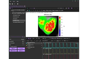
Cardiac Optical Mapping Data Anaysis using BV Workbench Version 4.5.1
Calcium Imaging of Cultured Cardiomyocyte Sheet
- Sample
- Cultured cardiomyocyte sheet
- Fluorescence Dye
- Calcium Dye (Cal-520)
- Imaging System
- MiCAM05-C35IR
- Pixels
- 636x360
- Frame Rate
- 200fps (5msec/frame)
- Data provided by
- Dr. Hiroko Izumi-Nakaseko, Department of Pharmacology, Faculty of Medicine, Toho University
High-Speed Calcium Imaging of Isolated Mouse Atrial Cardiomyocytes
- Sample
- Freshly isolated mouse atrial cardiomyocytes. Cells were plated on glass cover slips coated with laminin and used on the same day as isolation.
- Method
- Cells were paced by electrical field applied through a perfusion solution. Imaging was performed on Nikon DIAPHOT 300 microscope with 20X lens and SPECTRA-X Lumencor light source.
- Fluorescence Dye
- Calcium sensitive dye (Fluo-4 AM)
- Imaging System
- MiCAM05-ULTIMA
- Pixels
- 100x100
- Frame Rate
- 200fps (5.0msec/frame)
- Data provided by
- Dr. Roman Medvedev and Dr. Alexey Glukhov, Department of Medicine, University of Wisconsin-Madison
Monolayer of neonatal mouse ventricular myocyte stained with voltage sensitive dye

- Sample
- Monolayer of neonatal mouse ventricular myocyte. Sample was scratched with a needle tip at three locations (visible on the fluorescence pictures) to induce conduction blocks and meandering of the wavefront.
- Method
- Sample is paced locally at different frequencies using a glass microelectrode positioned at the edge of the sample
- Fluorescence Dye
- Voltage sensitive dye (Di-8-Anepps)
- Imaging System
- MiCAM03-N256
- Pixels
- 256x256
- Frame Rate
- 1,000fps (1.0msec/frame)
- Data provided by
- Dr. Jan Kucera and Dr. Ange Maguy, Department of Physiology, University of Bern
Calcium Imaging of Cultured Cardiomyocytes (Fluo-4)
The preparation was cultured rat cardiomyocytes stained with Fluo-4 and activity was recorded using MiCAM01 (old model) in 2002. The current model, MiCAM02 can significantly improve the data in terms of spatial resolution, sensitivity and S/N ratio.
Voltage-Sensitive Dye Imaging of Cultured Neonatal Rat Ventricular Cardiomyocytes
Sustained spiral wave reentry in a 3 day old monolayer disc of cultured neonatal rat ventricular cardiomyocytes (frequency ~ 5 Hz; diameter of the preparation = 8 mm). The preparation was stained with the voltage sensitive dye di-8-ANEPPS and activity was recorded at 1,000 frames/second using a custom-made tandem lens macroscope and an Ultima-L camera system.
- Data provided by
- Dr.Stephan Rohr, University of Bern
- High Speed Imaging of Membrane Potential using Voltage Sensitive Dye
- What is Voltage Sensitive Dye?
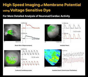
Voltage-sensitive dye changes its absorbance and fluorescence intensity when membrane potential changes in a stained brain or heart tissue.
By using voltage-sensitive dyes as chemical probes and capturing changes in light intensity with the use of a high-speed imaging device, it is possible to image in real time the activity of where, when, and how much excitation or inhibition occurred, in the brain and heart.
Product suitable for this kind of application
- High Speed Imaging System
- MiCAM03-N256
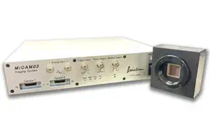
MiCAM03-N256 is a high-speed imaging system that captures and visualizes small changes in fluorescence intensity from biological samples stained with fluorescent probes, such as voltage sensitive dyes and calcium dyes.
- Spatial Resolution :
-
128x128 - 256x256 pixles
(32x32 pixels - 256x256 pixles with option) - Maximum Frame Rate :
- 1,000fps
(20,000 fps available with option) - Up to 2 camera heads can be used with completely synchronization.