BV Workbench is data acquisition/analysis software for the imaging systems (MiCAM05, MiCAM03, MiCAM ULTIMA, MiCAM02, etc.) manufactured by Brainvision. It also supports 16-bit grayscale TIFF files, and can read images and analyze data accquired by commercially available cameras.

- Data Acquisition/Analysis Software
-
BV Workbench
Main Features
- Can easily create various maps such as activation map and APD map with intuitive operation
- Peak detection and optimum value setting can be performed quickly with semi-automatic data analysis
Application
- Data analysis for brain and heart sample stained with fluorescence dye such as voltage sensitive dye / calcium dye
- Activation map analysis of tachycardia
Brochure Download
-
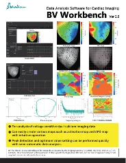
Data Analysis Software BV Workbench
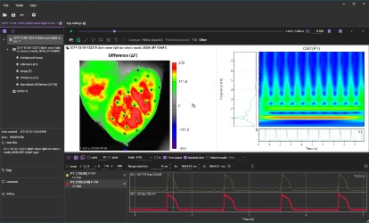
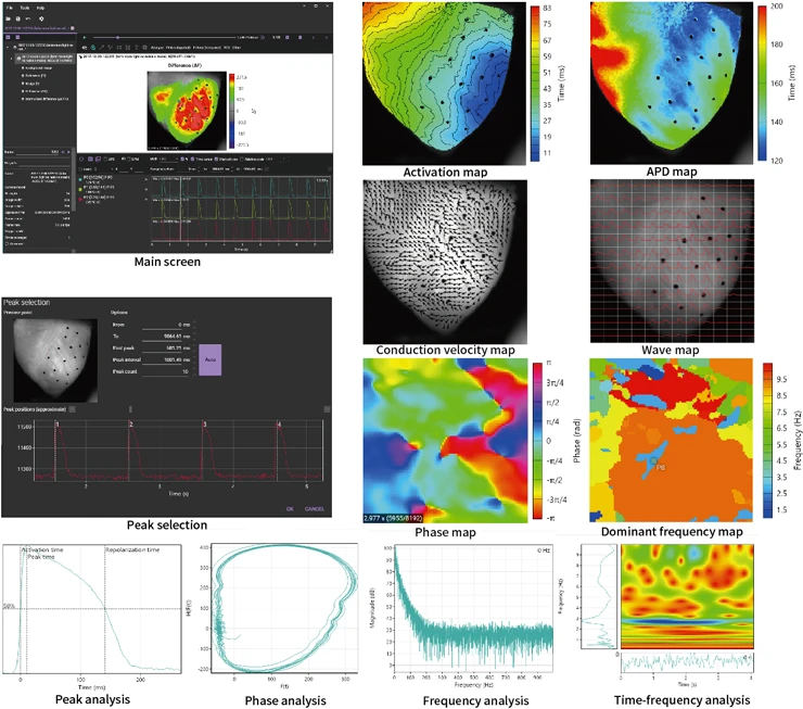
Main Screen
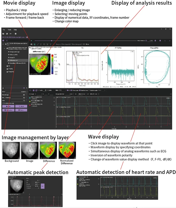
Data Analysis using BV Workbench - 1. Basic operation
Various Maps
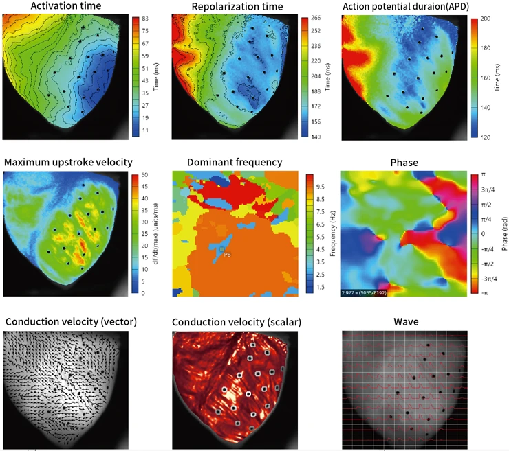
Action Potential
- Activation time
- Repolarization time
- Action potential duration (APD)
- Activation to peak time
- Peak to repolarization time
- Peak time
- Peak amplitude
- Decay tau
- Maximum upstroke velocity
Calcium transient
- Half rise time
- Half decay time
- Calcium transient duration (CaD)
- Half rise to peak time
- Peak to half decay time
- Peak time
- Peak amplitude
- Decay tau
- Maximum upstroke velocity
Data Analysis using BV Workbench - 4-1. Activation map
Data Analysis using BV Workbench - 4-2. Action potential duration (APD) map
Phase Mapping for Cardiac Optical Data
Activation map, Repolarization map and APD map by using BV Workbench
Analysis
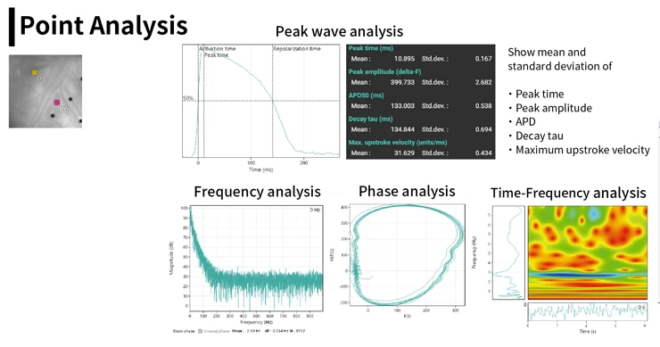
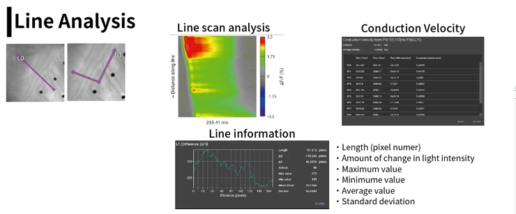
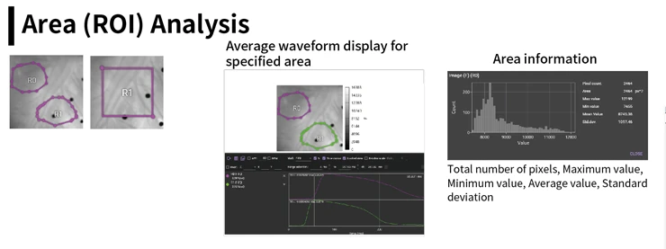
Data Analysis using BV Workbench - 2. Drawing tools and analysis (point, line, ROI)
Other Functions
Various Filters
| Undo | Undo last filter |
|---|---|
| Invert polarity | Reversal of image and wavelength polarity |
| Binning | Combine multiple pixels into one pixel |
| Brightness/Illumination correction | Brightness correction/Illumination light unevenness correction |
| Gaussian filter | Gaussian filter (noise removal) |
| Mean filter | Mean filter (noise removal) |
| Median filter | Median filter (noise removal) |
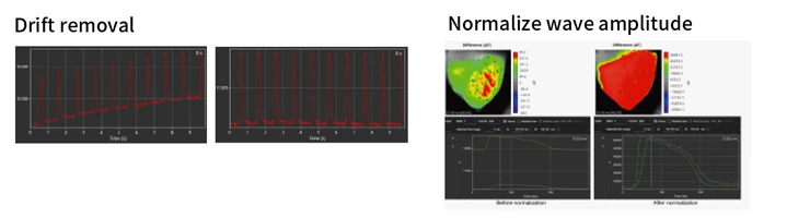
| Drift removal | Correct drift of baseline (bleach of fluorescence dye) |
| FIR Filter | FIR temporal filter (noise removal) |
| Dynamic range optimization | Optimize dynamic range |
| Normalize wave amplitude | Make maximum amplitude uniform |
| Filter batch | Apply multiple filters at once |
| Deinterleave frames | Divide frames of two wavelength imaging data into two data |
Other Functions
| Image align | Enlarge / reduce, rotate, move, and ovelay two images |
|---|---|
| Crop | Image crop |
| Arithmetic operation | Calculation processing between two data |
| Batch average | Manual averaging of multiple data |
| Create subset | Cut a part of data and save it as another data |
Data Export
- Image CSV (*.csv)
- Movie (*.avi)
- Image/Map data (*.png)
- Wave CSV (*.csv)
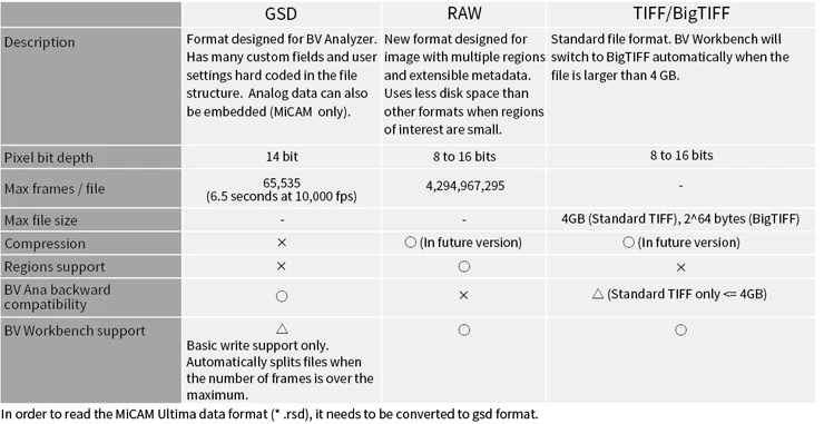
Supported Data Format
Software license
In order to use BV Workbench, you need to activate using a dedicated license (charged). This license is issued on a per computer basis using a hardware ID created based on information specific to an installed computer. You cannot authenticate with a license issued for another computer. Please contact us for details on how to obtain and set the license.
Download
The installer can be downloaded on the “Support" pages.
Recommended Specifications
| OS | Windows 10 64-bit |
|---|---|
| Software | .NET Framework 4.8 |
| RAM | 16GB or more |
| Graphic card | NVIDIA GPU Compute Capability 3.0 or more |
| Monitor | 1,280x1,028 dot or more |
- High Speed Imaging of Membrane Potential using Voltage Sensitive Dye
- What is Voltage Sensitive Dye?
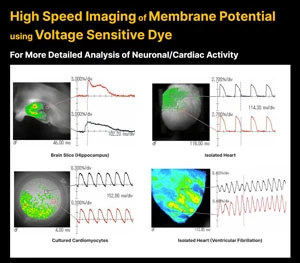
Voltage-sensitive dye changes its absorbance and fluorescence intensity when membrane potential changes in a stained brain or heart tissue.
By using voltage-sensitive dyes as chemical probes and capturing changes in light intensity with the use of a high-speed imaging device, it is possible to image in real time the activity of where, when, and how much excitation or inhibition occurred, in the brain and heart.