Why does the THT Mesoscope have high N.A even at low magnification?

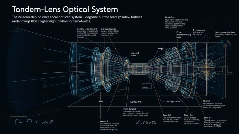
The Secret Behind Its 100-Fold Higher Light Collection Efficiency — "Tandem-Lens Optical System”
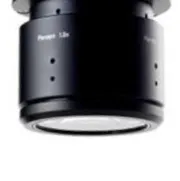
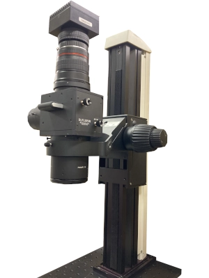 THT Mesoscope
The THT Mesoscope (THT series) is a macro fluorescence microscope developed
for wide-field imaging of biological activity in organs like the brain and
heart.
THT Mesoscope
The THT Mesoscope (THT series) is a macro fluorescence microscope developed
for wide-field imaging of biological activity in organs like the brain and
heart.
Its most remarkable feature is its ability to capture slight change in fluorescence with an extremely bright image and a high signal-to-noise (S/N) ratio, despite its low magnification range (approx. 0.19x to 6.3x).
In quantitative terms, the THT Mesoscope provides images that are about 30 times brighter at 1x total magnification and 10 times brighter at 2x, compared with conventional fluorescence stereo microscopes.
Furthermore, early research on the tandem-lens macroscope1), which formed the basis of its design, demonstrated a 100- to 700-fold advantage in fluorescence image brightness compared to commercially available microscope objectives of similar magnification.
This exceptional brightness and sensitivity stem from the innovative “tandem-lens optical design”, which overcomes a fundamental limitation of traditional low-magnification optics.
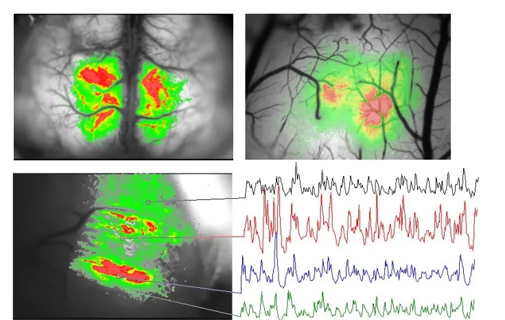
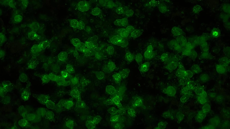
Biological activity acquired with THT Mesoscope (in vivo brain)
1. The “N.A Barrier” in Conventional Low-Magnification Microscopes
Why do images typically become darker when you reduce magnification to obtain a wider field of view? The fundamental reason lies in the low Numerical Aperture (N.A) of conventional objective lens.
While microscopes are optimized for high magnification and high NA to observe small objects, this excellent light-gathering efficiency is not maintained at the low magnifications required for wide-field imaging.
1-1. The Relationship Between NA and Brightness:
The brightness of a fluorescence image is primarily proportional to the square of the lens's NA 1). In the case of epi-illumination, the amount of excitation light also depends on the NA, meaning image brightness can even depend on the fourth power of the NA.
1-2. The limitation of available optics:
Commercially available low-magnification objective lenses have very low NA values. For instance, a 2.5x objective typically has an NA of 0.08, and a 1x objective has an NA of 0.04. Even for commercial stereo macroscopes, an NA of 0.03 at 6.3x was once considered "excellent." 1)
1-3. The Challenge of Weak Signals:
A low NA results in insufficient light collection efficiency, leading to dark images dominated by detector dark noise. Consequently, signal averaging becomes time-consuming, and the viability of experiments involving weak fluorescent signals—such as those using voltage-sensitive dyes (VSDs)—is limited by photobleaching and phototoxicity.
2. A Revolution in NA: The Tandem-Lens Design
To overcome the intrinsic low-NA problem, the THT Mesoscope applies the tandem-lens macroscope concept introduced in 1991.
A tandem-lens system consists of two infinity-corrected compound lenses (an
objective lens, L1, and an imaging lens, L2) placed front-to-front. In this
configuration, the magnification is simply determined by the ratio of the
focal lengths (FL) of the two lenses:
M = FLimaging/FLobjective 2)
2-1. Achieving High NA and Superior Light Throughput
The design of the tandem-lens system achieves a high NA of 0.4, significantly surpassing that of conventional low-magnification lenses. This improvement in NA provides a dramatic advantage in light collection efficiency.
Theoretical Advantage in Light Collection:
Compared to a conventional 2.5x, 0.08 NA objective, the theoretical
advantage in light collection efficiency, calculated by the ratio of the
squares of the NAs, is
(0.4)2/(0.08)2 ≈ 25x
Overall Light Throughput:
When measuring the total light throughput, which includes illumination efficiency, macroscopes have been shown to have a 300-fold advantage in standard configurations and up to a 700-fold advantage with small-field excitation.
This exceptional light throughput and high NA are the primary reasons why the THT Mesoscope can achieve extremely bright imaging at low magnifications.
2.2. Optimized for Camera-Based Imaging With Minimal Optical Loss
The THT Mesoscope employs a design philosophy thoroughly specialized for "camera-based imaging with the highest possible S/N ratio" by minimizing light loss.
Elimination of Light Loss Factors:
Elements that cause light loss, such as eyepieces for direct observation and zoom functions, have been intentionally excluded from the optical path.

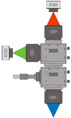
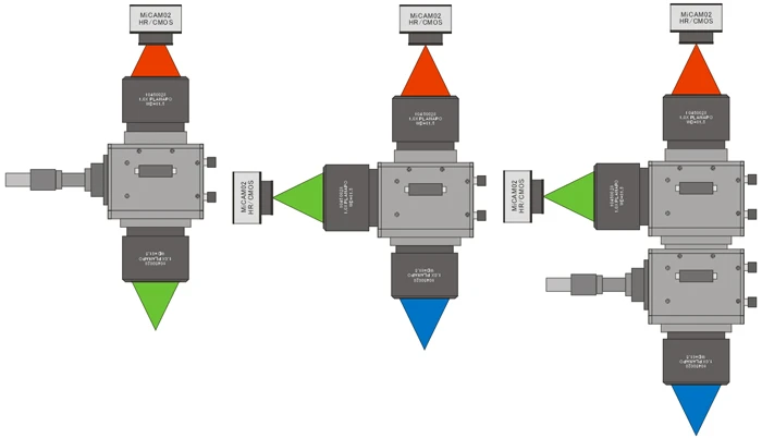
Diagram of THT Mesoscope (configuration for brain slice imaging)
Simplified optical path:
A pair of large-aperture objective lenses and custom large-diameter fluorescence filters create a straightforward, high-throughput light path that directs even slight change in fluorescence efficiently to the detector.
Large-aperture objective lense
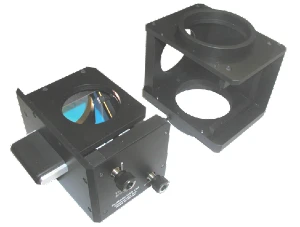
Custom large-diameter fluorescence filters
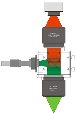
High-throughput light path
As a result, empirical measurements show approximately 30x brightness at 1x and 10x at 2x total magnification compared to conventional systems.
3. Decisive Advantages of High Brightness in Research
The THT Mesoscope's superior light collection efficiency and high S/N ratio offer several critical advantages for wide-field imaging research, extending beyond mere brightness.
Improved S/N Ratio and Experimental Longevity
A 300x gain in fluorescence intensity translates to a 17x improvement in S/N when shot noise dominates.
Moreover, with 20x higher collection efficiency, the excitation intensity can be reduced to 1/20th, allowing for much longer experiments before photobleaching or phototoxicity occur.
For example, while conventional systems could sustain only about four trials, researchers have reported obtaining data from 20 distinct stimulation conditions at comparable S/N using the THT Mesoscope.
Artifact Reduction with a Shallow Depth of Field
By achieving a high NA, the THT Mesoscope's design provides a 5 to 10 times shallower Depth of Field (DoF) than conventional low-magnification microscopes.
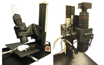
This shallow DoF is extremely useful for in vivo cortical imaging. When acquiring activity-dependent intrinsic signals via reflectance measurements, large blood vessels on the cortical surface often cause artifacts. With the THT Mesoscope, by focusing below the cortical surface (e.g., at a depth of 300 μm), artifacts from surface vessels can be eliminated, facilitating the imaging of cortical functional architecture. In contrast, with a standard macro lens, significant artifacts were observed even when focusing 600 μm below the surface.
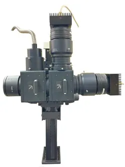
High Flexibility and Expandability
Leveraging its high light throughput, the THT Mesoscope offers the flexibility to accommodate diverse experimental needs.
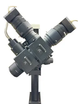
Simultaneous Multi-Wavelength Imaging:
By using the main body as a fluorescence beam splitter and connecting two cameras, simultaneous two-wavelength fluorescence imaging is possible.
Two-story building setup
Flexible Observation Angles:
An optional stand allows the optical axis to be rotated horizontally and tilted. This enables creative imaging setups, such as capturing the entire temporal lobe of the brain without having to tilt the animal sample.
Conclusion
The THT Mesoscope is exceptionally bright at low magnifications because it overcomes the fundamental limitation of conventional low-magnification optics by employing a tandem-lens system based on high-NA lenses.
Furthermore, by eliminating sources of light loss and adopting a simple optical path optimized for camera detection, the system maximizes the ability to detect weak fluorescence signals with a high S/N ratio.
This design significantly contributes to high-quality data acquisition in a wide range of applications, including voltage-sensitive dye imaging, in vivo wide-field calcium imaging (GCaMP), FRET, and GEVI imaging, enabling researchers to easily capture signals that were previously difficult to detect.
References:
1)
A tandem-lens epifluorescence macroscope: hundred-fold brightness
advantage for wide-field imaging
E H Ratzlaff 1, A Grinvald (1991)
2)
Functional Optical Imaging of Intrinsic Signals in Cerebral Cortex
L. E. Hallum, S. C. Chen, S. L. Cloherty, J. W. Morley, G. J. Suaning, N. H.
Lovell (1991)
Product information
- Fluorescence Mesoscope
- THT Series
The THT is a fluorescence widefield mesoscope developed for detecting low light fluorescence that is emitted from biological sample at a low magnicifation. This mesoscope has a very simple optical path combining two large-diameter objective lenses and a custom-made large fluorescent filter