Widefield Optical Imaging
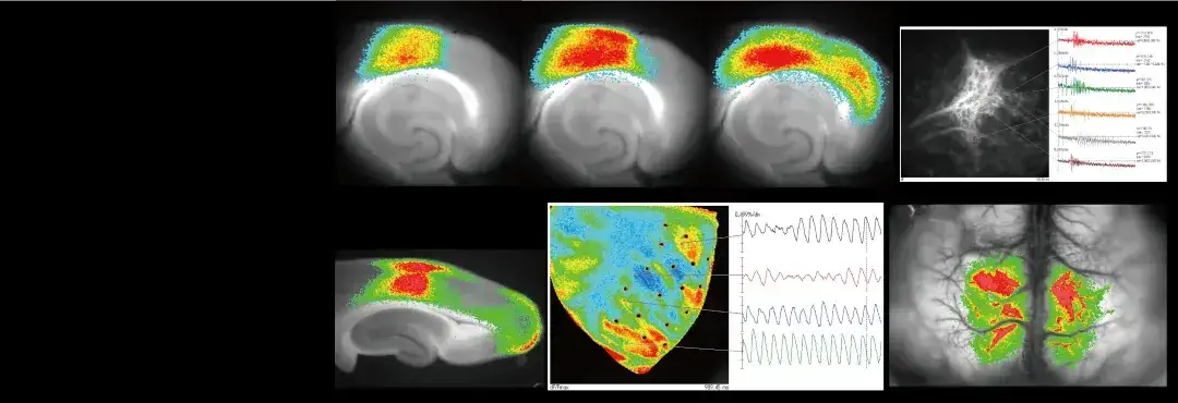


SciMedia specializes in complete turn-key, optical imaging systems for neuroscience and cardiac research.
1,000 fps at 256x256 pixels
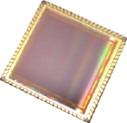
Original Large CMOS Image Sensor
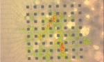
- Voltage Sensitive Dye
- Calcium Dye
- GCaMP
- GEVI
- Intrinsic Optical Signal
- and more...
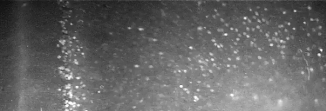
Products for High Speed Fluorescence Imaging
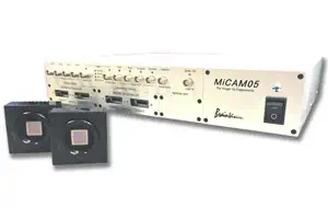
MiCAM05-N256
Multi-function, high-speed imaging system. Imaging speed of up to 1,923 fps with 256x256 pixels. Up to 4 cameras can be connected.
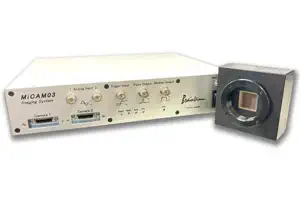
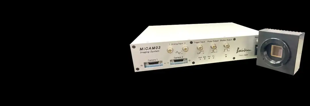
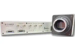
MiCAM03-N256
High-speed imaging system. Imaging speed of up to 1,000 fps with 256x256 pixels. Up to 2 cameras can be connected.
MiCAM03-C35IR
An ultra-sensitive, wide-field imaging system equipped with a large 35mm full-size CMOS sensor with 2,160x1,280 pixels.
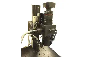
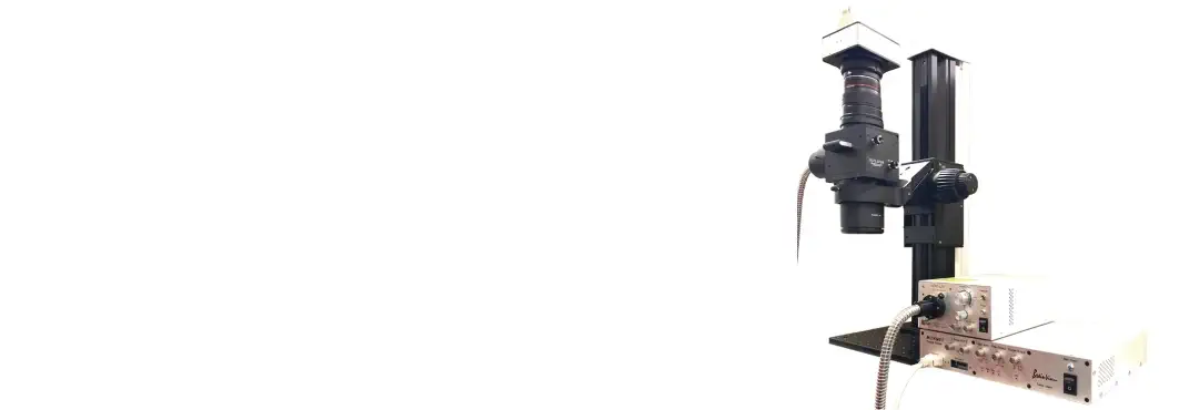
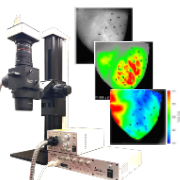
THT Series
A mesoscope developed for fluorescence imaging such as voltage sensitive dye imaging, calcium imaging and GCaMP imaging.

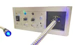
LEX9
An LED light source developed in pursuit of high brightness, high stability, and simple operability. Suitable for voltage sensitive dye imaging, calcium imaging and GCaMP imaging.
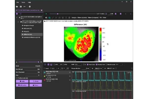
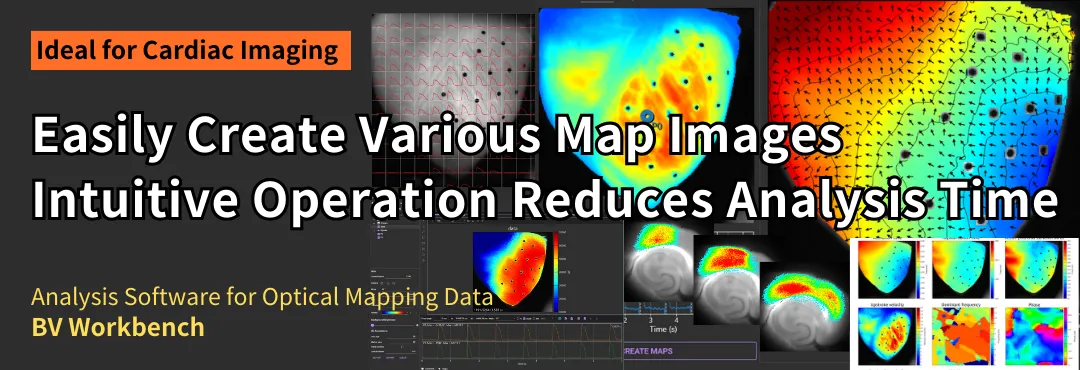
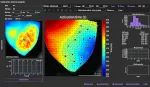
BV Workbench
Software developed for analysis of optical imaging data such as neural activity and cardiac activity. Various image analysis can be performed quickly.
Our Advantages

Imaging Applications
Other products
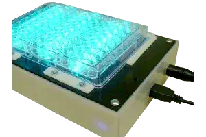
Cardiac Signal Acquisition System CIOS8
Activity of cardiomyocytes in a culture plate can be measured without invasion. Number of beats and contraction time are automatically detected
What's New?
-
-
-
-
 04/18/2022
Set up a new Twitter account. Please follow us @SciMediaLtd on Twitter -